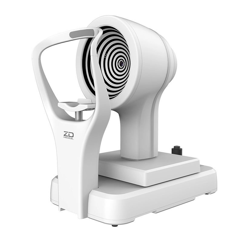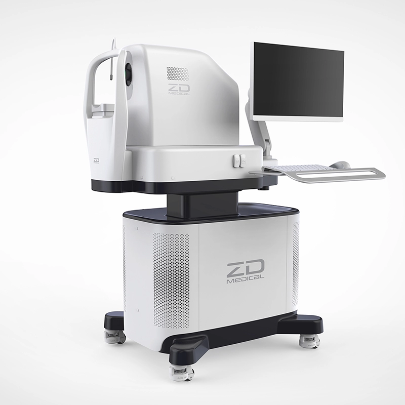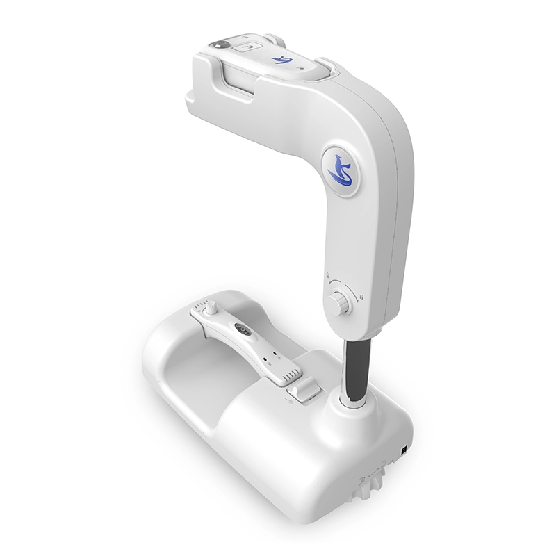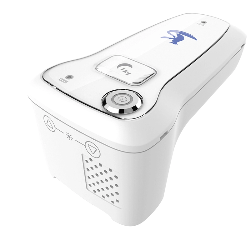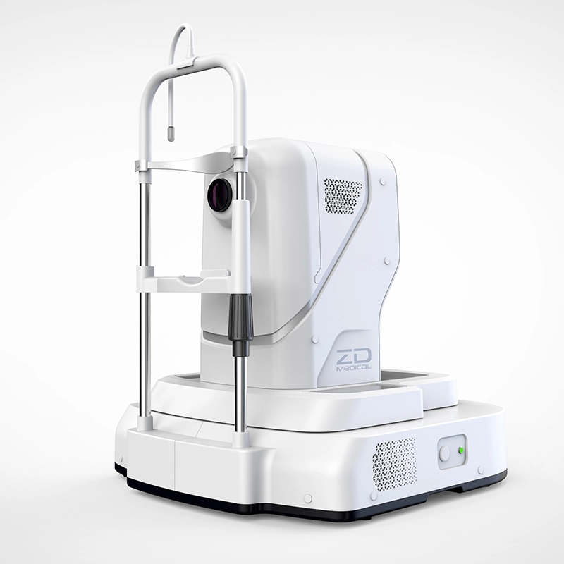
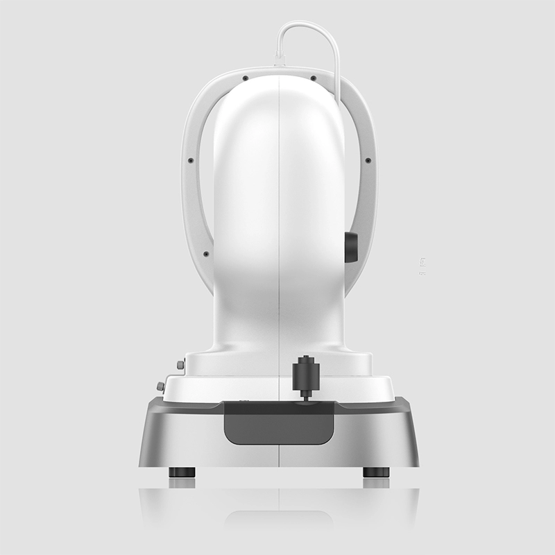
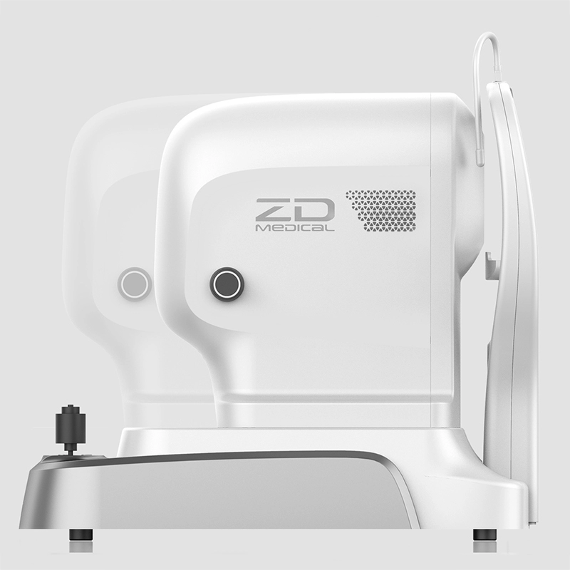
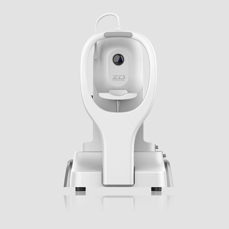
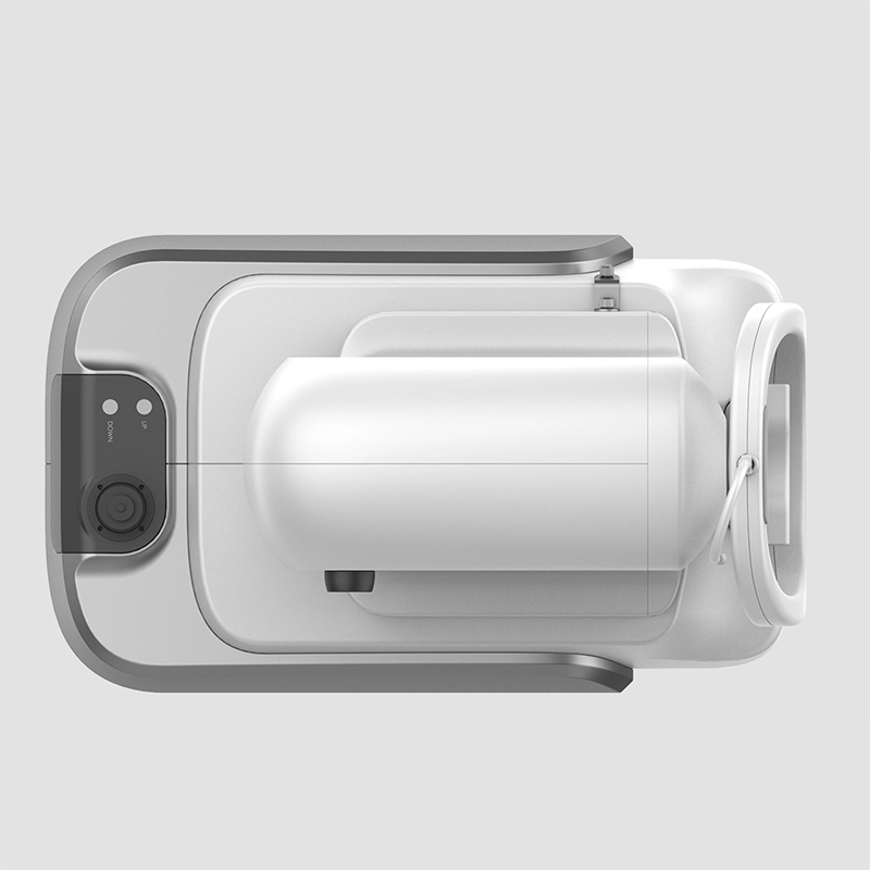
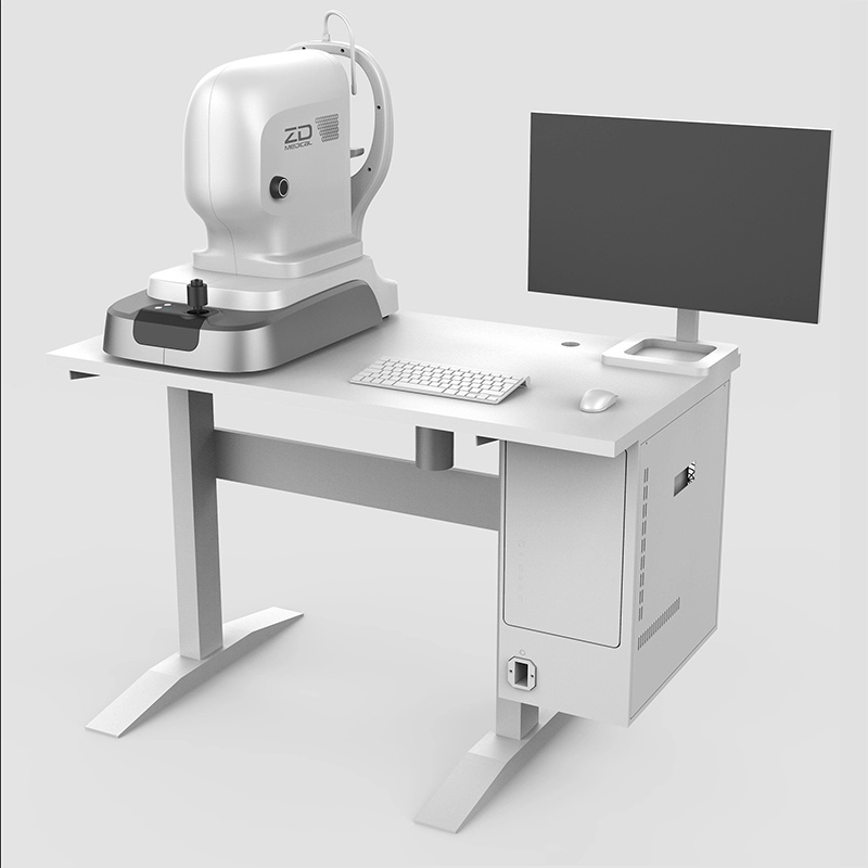
Optical Coherence Tomography OCTA2020
OCTA2020 from ZD Medical uses LSLO technology, with up to 2.65mm scan depth,and the lateral resolution of retinal fundus image is up to 5μm. Equipped with professional analysis software, OCTA2020 can obviously show the macular thickness under the macular thickness analysis model, which helps accurately identify clinical macular diseases.
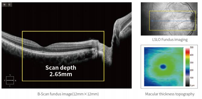
Deep to the Bottom of Fundus
The scan depth is up to 2.65mm, reaching to the choroid arid even the sclera. Fully display of all layers during one scan.
The scan depth (imaging depth) is one of the most important performance parameters in the OCT system. OCTA2020 has an obvious advantage in depth, that is, the choroid imaging is clearer without affecting the axial resolution. It has significant advantages for fundus diseases involving high depth and high resolution,such as choroid disease diagnosis.
Wide to the Edge of Vision
Large scanning range, clear macular area and optic disc area at a glance.
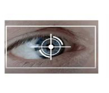
Auto Focus
OCT2020 can automatically complete the tracking of fundus and macula,detect and calibrate the central part of the pupil, detect and adjust the focus and fault position,and display retina layers of high-defition. The whole acquiring time is limited to 5 seconds,greatly saving the diagnosis time for clinicians.
Scan Mode
OCT2020 has a variety of scan modes,including area scan,HD one line scan and multilines scan.
Area scan∶512*64,range 6mm*6mm\range 12mm*12mm
HD one line scan: 1024*30,length 6mm;2048*30,length 12mm
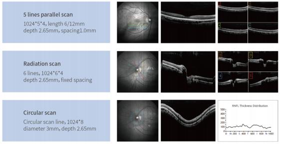
Mutilines scan:5 lines parallel scan, Radiation scan, Circular scan
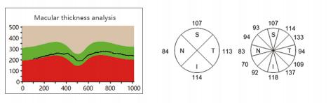
Glaucoma Analysis
Accurately measure the retinal thickness around the fovea and compare it with the age-elated normal data to analyze the deviation of patient's retinal thickness, which helps clinicians diagnose glaucoma.
Perform circular scan around the optic papillia, automatically quantify the optic nerve thickness, and compare the thickness with database, so as to examine the atrophy of the optic nerve as an early sign of glauco-
ma.
Follow-up, a More Efficient Patient Management
Accurate and rapid scan helps followup the disease changes,making diagnosis more efficient and easier.
OCT2020 can automatically record current scanning position of macular and eyeball and intelligently locate the previous scanning position in the later follow-up examination, to ensure that two scans are in the same position. Based on the trend analysis of retinal thickness in different stages, perform long-term follow-up examinations and trend analysis.
Anterior Segment Examination
Cornea scan
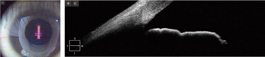
Clearly showing the cornea and iris
Precisely scanning the chamber corner, clearly showing the chamber corner structure
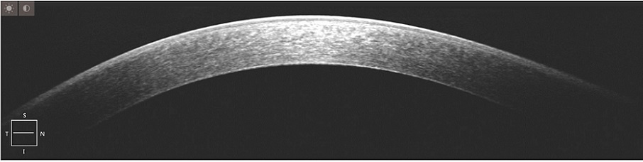
HOD cornea image, clearly showing the corneal epithelial layer,anterior elastic layer and coneal stroma
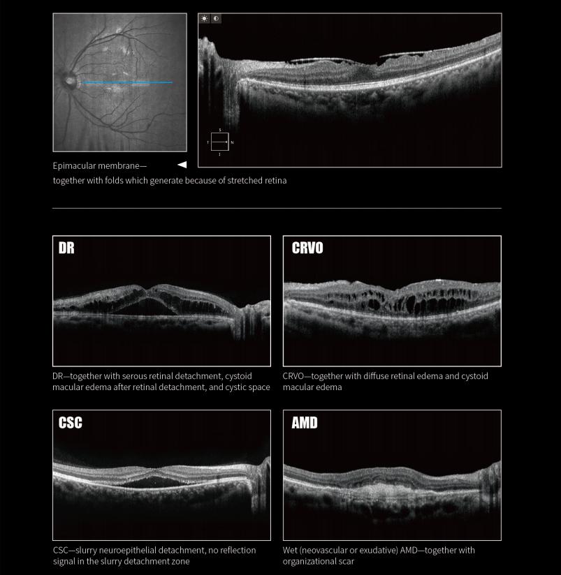
Fundus Imaging
Clear image collected by OCT 2020, help you diagnose the disease.
Technical Parameters
|
Measurement |
Axial resolution(in tissue): 5μm(in tissue) |
|
Horizontal resolution(in tissue): 10μm(in tissue) |
|
|
Scanning |
Maximum a scanning speed: 20KHz, tolerance±5% |
|
Maximum scanning depth≥2.65mm(in tissue), tolerance±3% |
|
|
Maximum scanning range: 12mm × 12mm, tolerance±5% |
|
|
Light Source |
Central wavelength: 843nm |
|
Light power≤740μm(at the Cornea) |
|
|
Refractive compensation range: -20D~+20D |
|
|
Fundus Image |
Method: LSLO |
|
Central wavelength: 780nm |
|
|
Range: 42.0° × 42.0° |
|
|
B-scan |
Area scan: 512*64,r ange 6mm × 6mm 512*64, range 12mm × 12mm |
|
HD one line scan:1024*30,length 6mm; 2048*30,length 12mm |
|
|
Multi lines scan: 5 lines parallel scan, radiation scan, circular scan |
|
|
Other Functions |
Follow-up, Auto focus, Auto reference arm, Automatic segmentation and-manual segmentation, Anterior, Pseudo color, Glaucoma analysis, Macular thickness analysis(macular topographic map), Automatic identification for fovea centralis, Internal fixation target and external fixation target, RNFL clock hours, Eye-tracking, Cataract image enhancement、Common case library reference、Patient data management、Angle measurement、High-precision thickness measurement (can be zoomed in)、Multiple overlay optimization.
|
|
PC and Printer |
Hard disc: 2T |
|
CPU: I7-8700 |
|
|
GPU: RTX2060 6G |
|
|
RAM: DDR4 16G |
|
|
Display: 24-inch LCD screen |
Certificates:

Subscribe to our weekly newsletter and receive exclusive offers on products you love!
 X
X


 Gold Supplier